What Is the PERC Rule (Pulmonary Embolism Rule-out Criteria)?
The PERC rule calculator (Pulmonary Embolism Rule-out Criteria) represents one of the most critical clinical decision support tools in modern emergency medicine and acute internal medicine triage. Formulated and validated by Dr. Jeffrey A. Kline, MD and colleagues in 2004, the PERC criteria were created to solve a widespread crisis in clinical medicine: the rampant over-utilization of D-dimer blood assays and computed tomography pulmonary angiography (CTPA) in patients with an extraordinarily low likelihood of venous thromboembolism.
When an emergency patient presents with acute pleuritic chest pain, dyspnea, or syncope, pulmonary embolism (PE) frequently enters the differential diagnosis. Historically, fear of missing a potentially fatal vascular clot compelled emergency physicians to order D-dimer assays indiscriminately. However, because D-dimer is an acute-phase reactant with high sensitivity but notoriously low specificity, up to 70% of older adults or hospitalized patients return false-positive results due to trivial inflammation, recent sprains, or age. These false-positive laboratory values cascade into mandatory CTPA scans, subjecting young and elderly patients alike to substantial ionizing radiation, risk of iodinated contrast-induced nephropathy, and accidental detection of clinically inconsequential subsegmental clots.
The perc score calculator resolves this dilemma by serving as an objective "stopping rule." Rooted in rigorous threshold decision analysis, the perc calculator provides emergency clinicians with legal and scientific backing to rule out pulmonary embolism clinically—without drawing blood, without radiation, and without anticoagulant exposure—provided the patient satisfies all eight high-sensitivity criteria. Whether used alongside the Wells Score Calculator or integrated into standard emergency room triage pathways, the PERC rule protects patients from unnecessary iatrogenic harm.
The 8 PERC Criteria: HAD CLOTS Mnemonic & Pathophysiological Rationale
To ensure rapid recall during high-stress emergency resuscitations, the eight components of the perc criteria calculator are commonly remembered via the clinical mnemonic HAD CLOTS. Each variable reflects a distinct pathophysiological pillar of Virchow's triad (endothelial injury, venous stasis, hypercoagulability) or cardiopulmonary strain:
| Mnemonic | PERC Criterion | Clinical Cutoff | Pathophysiological Relevance in PE Triage |
|---|---|---|---|
| H | Heart Rate (Tachycardia) | ≥ 100 Beats / Minute | Resting sinus tachycardia represents early right ventricular strain and compensatory neurohormonal activation to maintain cardiac output against acute pulmonary vascular resistance. |
| A | Age Threshold | ≥ 50 Years | Baseline incidence of venous thromboembolism increases exponentially after age 50 due to cumulative venous valvular incompetence, endothelial atheroma, and prothrombotic fibrinogen elevation. |
| D | DVT or PE Prior History | Documented Prior VTE | A personal history of deep vein thrombosis or pulmonary embolism indicates intrinsic hypercoagulability (thrombophilia, residual post-thrombotic syndrome), carrying a persistent risk multiplier. |
| C | Coughing Blood (Hemoptysis) | Any Frank Blood | Microvascular pulmonary infarction occurs when peripheral emboli occlude distal pulmonary arteries, causing alveolar hemorrhage and bronchial mucosal extravasation. |
| L | Leg Swelling (Unilateral) | Asymmetric Edema | Asymmetric calf enlargement or deep venous tenderness indicates active acute lower extremity DVT, the anatomical source of over 90% of all pulmonary emboli. |
| O | Oxygen Saturation (SpO2) | < 95% on Room Air | Ventilation-perfusion (V/Q) mismatch caused by occluded vascular beds and reflex bronchoconstriction lowers ambient arterial oxygen tension below 95%. |
| T | Trauma or Recent Surgery | Within Past 4 Weeks | Major procedures under general anesthesia, pelvic fractures, or lower extremity casting induce endothelial trauma and prolonged venous stasis. |
| S | Sex Hormones / Estrogen Use | Active Oral / Transdermal | Exogenous estrogens (combined oral contraceptive pills, hormone replacement therapy) increase hepatic synthesis of clotting factors (II, VII, VIII, X) while decreasing antithrombin III. |
Visual Architecture: The Emergency Pulmonary Embolism Diagnostic Algorithm
A frequent error among clinicians is attempting to apply the perc rule for pe calculator in isolation. The PERC criteria are not a standalone diagnostic test; they function as a refined second-stage filter within a structured, two-step clinical algorithm. As visualized in the emergency algorithm below, pre-test probability assessment must always precede PERC evaluation:
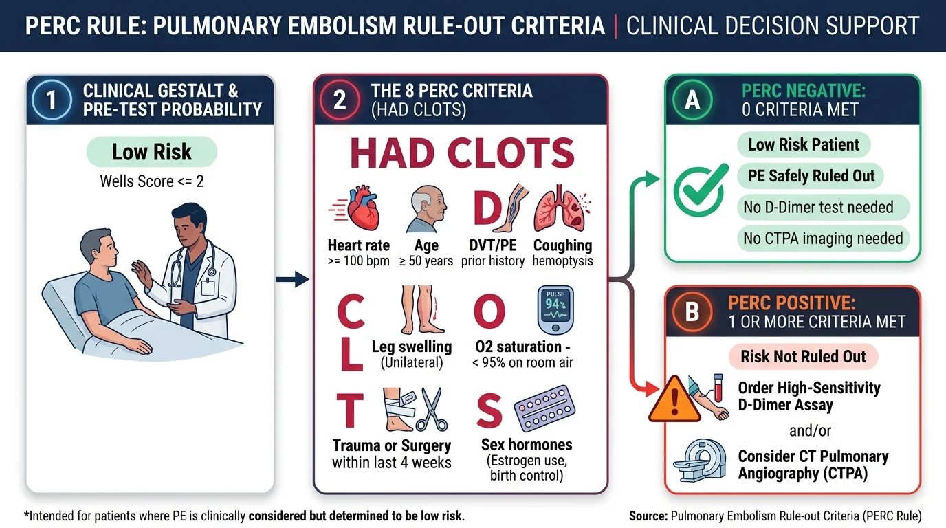
As demonstrated in the clinical flowchart above, patient management flows through three distinct checkpoints:
- Checkpoint 1 — Low-Risk Gestalt Filter: The clinician evaluates baseline clinical probability. If the patient is categorized as Low Risk (physician gestalt < 15% or Wells Score ≤ 2.0), proceed directly to Step 2. If the patient is moderate or high risk, PERC is invalid; proceed directly to D-dimer or CTPA.
- Checkpoint 2 — The 8 PERC Criteria (HAD CLOTS): If all 8 criteria are absent (0 points), the patient is PERC Negative. PE is ruled out; testing stops immediately. If 1 or more criteria are present, the patient is PERC Positive.
- Checkpoint 3 — Targeted Objective Testing: For PERC-positive patients, order a high-sensitivity D-dimer assay. In adults older than 50, apply the age-adjusted cutoff (Age × 10 μg/L) to prevent false-positive CT referrals. If D-dimer is positive, perform multidetector CTPA imaging.
The Test-Threshold Concept: Why PERC Negative (< 1.8% Risk) Avoids Testing
The mathematical foundation of the perc calculator mdcalc alternative is based on the Pauker-Kassirer Test-Treatment Threshold Model. In clinical epidemiology, every diagnostic test carries an inherent risk of iatrogenic complications. Diagnostic testing is only rational when the pre-test probability of a disease is higher than the test threshold ($T_{\text{test}}$):
For pulmonary embolism, the testing threshold is calculated based on the mortality and morbidity of CTPA and anticoagulant therapy:
- Contrast-Induced Acute Kidney Injury & Anaphylaxis: Iodinated contrast carries a 1% to 2% risk of renal impairment, particularly in patients with baseline borderline eGFR levels or diabetic nephropathy.
- Radiation-Induced Carcinogenesis: A single chest CT delivers approximately 7 to 10 mSv of ionizing radiation, conveying a tangible lifetime attributable risk of breast and lung malignancy, particularly in young female patients.
- False-Positive D-Dimer Cascades: Low-risk patients with mildly elevated D-dimer who undergo CTPA frequently have indeterminate or false-positive subsegmental findings. This can result in 3 to 6 months of unnecessary oral anticoagulation, exposing them to major hemorrhage risks calculated by our HAS-BLED Score Calculator.
Extensive clinical trial data demonstrates that the test threshold for PE is approximately 1.8% to 2.0%. If a clinical decision rule can prove that a patient's risk is below 1.8%, the harm caused by performing a CT scan or D-dimer test mathematically exceeds the harm of the disease itself. Because a PERC Negative score (0/8 criteria) guarantees a post-test PE probability between 0.5% and 1.4%, ordering further testing violates evidence-based medicine principles.
Wells Score vs. PERC Rule: Crucial Clinical Differences & Sequencing
Medical students, residents, and nurse practitioners frequently confuse how the Wells Score for PE and the perc score pe calculator interact. The table below outlines their distinct roles and clinical hierarchy:
| Comparison Dimension | Wells Score for PE | PERC Rule (Pulmonary Embolism Rule-out Criteria) |
|---|---|---|
| Primary Purpose | Risk stratification into prognostic tiers (Unlikely vs. Likely; Low, Mod, High). | Rule-out filter to eliminate blood tests and radiation in low-risk patients. |
| When Applied? | First-line evaluation for all patients with suspected acute PE. | Second-line filter, applied only if Wells score is ≤ 2.0 (Low Risk). |
| Number of Criteria | 7 weighted clinical criteria (from 1.0 to 3.0 points each). | 8 unweighted binary criteria (all criteria must be absent). |
| Can it Eliminate D-Dimer? | No. Low-risk Wells patients (≤ 4.0 pts) still require a D-dimer test. | Yes. PERC Negative patients do not require any D-dimer test. |
| Applicable in High Risk? | Yes. Identifies patients requiring immediate CTPA without D-dimer. | Strictly Contraindicated. Never use in moderate or high-risk patients. |
| Clinical Sensitivity | ~75% to 85% across all tiers. | > 97% to 99% sensitivity in low-risk emergency cohorts. |
Clinical Validation Evidence: The PROPER Trial & Multicenter Trials
The safety and efficacy of the perc online calculator have been validated across some of the largest multicenter prospective cohorts and randomized controlled trials in emergency cardiology:
1. The Derivation & Initial Multicenter Validation (Kline et al., 2004 & 2008)
In the original derivation study published in the Journal of Thrombosis and Haemostasis, Dr. Kline and colleagues evaluated 1,872 emergency department patients. They established that among patients with a low pre-test gestalt probability (< 15%), meeting all 8 PERC criteria yielded a negative predictive value (NPV) of 99.3%, reducing false-negative missed emboli to 0.7%. A subsequent multicenter validation of 8,138 patients in 2008 confirmed a negative likelihood ratio of 0.17.
2. The PROPER Randomized Controlled Trial (JAMA 2018)
To provide definitive level-1 evidence, Freund et al. conducted the PROPER trial (Pulmonary Embolism Rule-Out Criteria Trial), published in JAMA in 2018. This cluster-randomized, crossover non-inferiority study enrolled 1,918 emergency department patients across 14 hospital centers. Key findings included:
- Non-Inferior Safety: Failure rate (subsequent thromboembolism at 3 months) was only 0.15% in the PERC group vs. 1.15% in the control group (proving non-inferiority).
- Massive Reduction in CT Scans: Use of chest CTPA was reduced by 36% (from 23% in the control arm to 13% in the PERC arm).
- Reduced Hospital Stays: Average emergency department length of stay decreased significantly, saving health systems millions of dollars and preventing unwarranted radiation.
PERC Positive Diagnostic Pathways & Age-Adjusted D-Dimer Integration
What should you do when a patient is PERC Positive? If even a single criterion is triggered (for instance, a 54-year-old female whose only positive factor is age ≥ 50), the PERC rule cannot be used to rule out pulmonary embolism. The physician must proceed to laboratory evaluation with a high-sensitivity quantitative D-dimer assay.
To prevent over-testing in older adults who fail PERC solely based on the age criterion, guidelines from the European Society of Cardiology (ESC) and American College of Emergency Physicians (ACEP) mandate using the Age-Adjusted D-Dimer Cutoff:
- Age ≤ 50 Years: Standard fixed cutoff of 500 μg/L (0.50 μg/mL FEU).
- Age > 50 Years: Adjusted cutoff calculated as: $$\text{D-Dimer Cutoff} = \text{Age} \times 10 \;\mu\text{g/L}$$
For example, a 68-year-old patient who fails PERC has an age-adjusted D-dimer threshold of 680 μg/L. If their measured D-dimer is 590 μg/L, PE is safely ruled out despite failing PERC, preventing an unwarranted CTPA scan.
Clinical Worked Scenarios: PERC Rule in Emergency Practice
Young Adult with Pleuritic Chest Pain: PERC Negative
Patient Presentation: A 26-year-old male presents to the ED with acute right-sided chest pain that sharpens upon deep inspiration. Symptoms began after lifting heavy weights. Vitals: BP 124/78 mmHg, HR 72 bpm, SpO2 99% on room air. He is non-obese, does not take medications, has no swelling, no hemoptysis, and no history of surgery or VTE.
Clinical Decision: Because gestalt is low risk and all 8 PERC criteria are absent, pulmonary embolism is safely ruled out. The clinician withholds D-dimer and CTPA, diagnosing musculoskeletal chest wall strain. The patient is discharged safely with anti-inflammatory analgesia.
Patient on Oral Contraceptives with Tachycardia: PERC Positive
Patient Presentation: A 31-year-old female presents with subacute mild dyspnea and palpitations. She takes combined oral contraceptive pills for polycystic ovarian syndrome. Triage vitals: BP 118/72 mmHg, HR 106 bpm (sinus tachycardia), SpO2 96% on room air. Lungs are clear, calves are non-tender, no hemoptysis, no prior surgery.
Clinical Decision: Although the patient is young and pre-test risk is low, she triggers two PERC criteria (Heart rate ≥ 100 bpm and exogenous estrogen use). PERC rule fails to exclude PE. The ED team orders a quantitative D-dimer assay. If D-dimer is < 500 μg/L, PE is ruled out. If elevated, CTPA imaging is performed.
When the PERC Rule MUST NOT Be Used: Critical Contraindications
Applying the perc calculator in inappropriate clinical contexts can result in catastrophic misdiagnoses. Emergency physicians and hospitalists must avoid using PERC under the following five conditions:
- Moderate or High Pre-Test Risk (Wells > 2.0): If your clinical intuition or scoring models suggest moderate or high probability, PERC is strictly invalid. Even with 0 criteria, residual risk in high-risk patients remains between 6% and 20%, which is unacceptably lethal.
- Hemodynamic Instability: Any patient with sustained hypotension (systolic blood pressure < 90 mmHg), circulatory shock, altered mental status, or right ventricular collapse requires immediate emergency resuscitation, bedside echocardiography, and emergent CTPA or empiric unfractionated heparin.
- Pregnancy: In pregnant women, baseline hypercoagulability, physiologic tachycardia, and elevated D-dimer levels invalidate PERC. Suspected PE in pregnancy requires specialized pregnancy-adapted algorithms (such as the YEARS algorithm or compression duplex ultrasonography before V/Q imaging).
- Active Malignancy with Unexplained Dyspnea: While active cancer is not an explicit PERC variable, it significantly increases baseline pre-test probability above 15%, moving patients out of the low-risk category.
- Severe Hypoxemia Requiring Supplemental Oxygen: If a patient requires supplemental oxygen to maintain SpO2 ≥ 95%, the true room air oxygen saturation is unknown, and the hypoxemia criterion must be considered positive.
Frequently Asked Questions About the PERC Rule Calculator
What is the PERC Rule and what does it calculate?
The PERC Rule (Pulmonary Embolism Rule-out Criteria) is an evidence-based clinical decision instrument developed by Dr. Jeffrey Kline in 2004. It evaluates eight high-sensitivity objective criteria in patients who are already judged to be at low pre-test risk (< 15% probability or Wells score ≤ 2.0) for pulmonary embolism. If all 8 criteria are negative, the patient's probability of PE drops below 1.8%, allowing safe rule-out without blood tests (D-dimer) or diagnostic imaging (CTPA).
What are the 8 PERC criteria (HAD CLOTS)?
The 8 PERC criteria are represented by the mnemonic HAD CLOTS: (1) Hormone / estrogen use, (2) Age ≥ 50 years, (3) DVT or PE prior documented history, (4) Coughing blood / hemoptysis, (5) Leg swelling / unilateral edema, (6) Oxygen saturation (SpO2) < 95% on room air, (7) Tachycardia / heart rate ≥ 100 bpm, and (8) Surgery or trauma requiring hospitalization within the past 4 weeks.
Can the PERC Rule be applied to moderate or high-risk patients?
No. The PERC Rule must NEVER be applied to patients categorized as moderate or high risk by clinical gestalt or a Wells PE score > 2.0. In higher-risk cohorts, a negative PERC score still leaves a substantial residual post-test probability of PE (> 5%–15%), which is unacceptably dangerous. Higher-risk presentations mandate immediate objective evaluation with D-dimer or CT pulmonary angiography.
What does a 'PERC Negative' result mean?
A 'PERC Negative' result occurs when a low-risk patient meets zero (0 out of 8) criteria. This means the post-test probability of PE is below 1.8%, which is beneath the Pauker-Kassirer test threshold. In this scenario, the risk of contrast-induced nephropathy, radiation exposure, and false-positive D-dimer cascades exceeds the risk of a missed PE. No D-dimer test and no CT scan are indicated; the patient can be safely evaluated for alternative non-thrombotic diagnoses.
What should be done if a patient is 'PERC Positive' (score ≥ 1)?
A patient is PERC Positive if even a single criterion (1 or more) is present. In this case, PE cannot be ruled out clinically. The clinician should proceed to the next diagnostic step: ordering a quantitative high-sensitivity D-dimer assay (or applying age-adjusted D-dimer cutoffs for patients aged > 50). If the D-dimer is elevated, CT Pulmonary Angiography (CTPA) or V/Q scanning is indicated.
How does the PERC Rule differ from the Wells Score for PE?
The Wells Score for PE is a pre-test probability stratification tool that calculates a point score (0 to 12.5) to divide patients into low, intermediate, or high risk (or 'PE Unlikely' vs. 'PE Likely'). The PERC Rule is a secondary rule-out filter used exclusively within the 'Low Risk' or 'PE Unlikely' subgroup to identify patients who can bypass D-dimer testing altogether. Wells categorizes risk; PERC eliminates unnecessary testing.
Peer-Reviewed References & Scientific Consensus
- Kline JA, Mitchell AM, Kabrhel C, Richman PB, Courtney DM. Clinical criteria to prevent unnecessary diagnostic testing in emergency department patients with suspected pulmonary embolism. Journal of Thrombosis and Haemostasis. 2004;2(8):1247–1255. doi:10.1111/j.1538-7836.2004.00790.x.
- Kline JA, Courtney DM, Kabrhel C, et al. Prospective multicenter evaluation of the pulmonary embolism rule-out criteria. Journal of Thrombosis and Haemostasis. 2008;6(5):772–780. doi:10.1111/j.1538-7836.2008.02944.x.
- Freund Y, Cachanado M, Aubry A, et al. Effect of the Pulmonary Embolism Rule-Out Criteria on Subsequent Thromboembolic Events Among Low-Risk Emergency Department Patients: The PROPER Randomized Clinical Trial. JAMA. 2018;319(6):559–566. doi:10.1001/jama.2017.21971.
- Konstantinides SV, Meyer G, Becattini C, et al. 2019 ESC Guidelines for the diagnosis and management of acute pulmonary embolism developed in collaboration with the European Respiratory Society (ERS). European Heart Journal. 2020;41(4):543–603. doi:10.1093/eurheartj/ehz405.
- Righini M, Van Es J, Den Exter PL, et al. Age-adjusted D-dimer cutoff levels to rule out pulmonary embolism: the ADJUST-PE study. JAMA. 2014;311(11):1117–1124. doi:10.1001/jama.2014.2135.