6-Step Analysis · Winter's Formula · Tic-Tac-Toe Visual

ABG Calculator:
Complete Arterial
Blood Gas Interpretation

The most comprehensive free ABG interpretation calculator available. Enter pH, PaCO₂, and HCO₃⁻ to instantly get primary disorder classification, compensation adequacy using Winter's formula, anion gap with albumin correction, P/F ratio, A-a gradient, base excess analysis, tic-tac-toe visual, and a full step-by-step breakdown. Built for nurses, respiratory therapists, medical students, and ICU clinicians.

What Makes This ABG Calculator Different From All Others

Most ABG calculators just label the disorder. Ours gives you everything in one place: primary disorder, compensation status (with formula), anion gap + albumin correction + delta-delta, P/F ratio, A-a gradient, base excess, Henderson-Hasselbalch consistency check, tic-tac-toe visual, 6-step teaching breakdown, and a 5-case practice quiz — all free, no login.

Normal ABG Values (Adult)
pH 7.35–7.45 PaCO₂ 35–45 mmHg HCO₃⁻ 22–26 mEq/L PaO₂ 80–100 mmHg BE −2 to +2
6
Interpretation Steps
4
Acid-Base Disorders
8+
Calculated Metrics
5
Practice Cases

⚠️ For educational use only. This tool supports learning and double-checking — it does not replace clinical judgment, senior review, or patient assessment. Always verify ABG interpretation in the context of the patient's full clinical picture.

ABG Interpreter

Clinical Formulas

Normal: 7.35–7.45

Normal: 35–45 mmHg

Normal: 22–26 mEq/L · Calculated value

Normal: 80–100 mmHg

Room air = 21%

For A-a gradient

Normal: −2 to +2

Normal: 4.0 g/dL

pH
PaCO₂
HCO₃⁻
PaO₂
P/F Ratio
Enter PaO₂ + FiO₂
Corr. AG
Enter Na⁺ + Cl⁻
Compensation Analysis
Formula Applied
Expected Value
Measured Value
Assessment
Tic-Tac-Toe ABG Visual
pH
PaCO₂
HCO₃⁻
Primary Disorder
Compensation
Normal
📋 Step-by-Step ABG Calculation Walkthrough

ABG interpretation is a clinical skill — this tool assists with calculations and teaching, not clinical decision-making. Mixed disorders, artefactual samples, and patient-specific factors can all affect interpretation. Always correlate with the patient's history, examination, and trend.

Normal ABG Values: What Does a Normal Arterial Blood Gas Look Like?

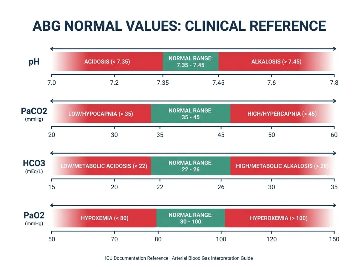
Understanding what a normal ABG looks like is the foundation of all interpretation. The body maintains these parameters within narrow ranges because even small deviations can signal significant pathology. What is a normal ABG value? The following reference ranges apply to healthy adults breathing room air at sea level.

Parameter US (mmHg / mEq/L) SI (kPa / mmol/L) What It Measures
pH7.35 – 7.457.35 – 7.45Overall acid-base status
PaCO₂35 – 45 mmHg4.7 – 6.0 kPaRespiratory component (CO₂ level)
HCO₃⁻ (calculated)22 – 26 mEq/L22 – 26 mmol/LMetabolic component (renal buffer)
PaO₂80 – 100 mmHg10.7 – 13.3 kPaOxygen level in arterial blood
SaO₂94 – 100%94 – 100%Haemoglobin oxygen saturation
Base Excess (BE)−2 to +2 mEq/L−2 to +2 mmol/LMetabolic buffering capacity
Anion Gap8 – 12 mEq/L8 – 12 mmol/LUnmeasured anions in plasma

Which ABG values are measured vs calculated? Only three values are directly measured by the blood gas analyser: pH (via a glass electrode), PaCO₂ (Severinghaus CO₂ electrode), and PaO₂ (Clark polarographic electrode). Bicarbonate (HCO₃⁻), base excess, and standard bicarbonate are all calculated by the machine using the Henderson-Hasselbalch equation. This is why ABG interpretation guidelines recommend checking consistency — if the calculated pH from the Henderson-Hasselbalch equation differs by more than ±0.05 from the measured pH, a sampling or processing error is likely.

The Four Primary ABG Acid-Base Disorders

Every ABG abnormality falls into one of four primary categories, or a combination (mixed disorder). Understanding what causes each disorder is as important as recognising the pattern from the numbers.

DisorderpHPaCO₂HCO₃⁻Primary Cause
Resp Acidosis↓ Low↑ High↑ (compensation)Hypoventilation — CO₂ retention
Resp Alkalosis↑ High↓ Low↓ (compensation)Hyperventilation — CO₂ loss
Met Acidosis↓ Low↓ (compensation)↓ LowHCO₃⁻ loss or acid gain
Met Alkalosis↑ High↑ (compensation)↑ HighH⁺ loss or HCO₃⁻ gain
🌬️
What Causes Respiratory Acidosis?

Any condition that reduces alveolar ventilation, causing CO₂ to accumulate. Common causes: COPD exacerbation, severe asthma, opioid or sedative overdose, chest wall injury, neuromuscular disease (Guillain-Barré, myasthenia gravis), sleep apnoea, and inadequate mechanical ventilation settings.

💨
What Causes Respiratory Alkalosis?

Any stimulus to hyperventilation causing excessive CO₂ loss. Common causes: anxiety/panic attacks, pain, hypoxia-driven hyperventilation (PE, pneumonia, high altitude), fever, sepsis, hepatic encephalopathy, pregnancy (progesterone stimulates respiratory centre), and over-ventilation on a ventilator.

🧪
What Causes Metabolic Acidosis?

Either acid accumulation or bicarbonate loss. Use MUDPILES for high anion gap causes: Methanol, Uraemia, DKA, Propylene glycol/Paracetamol, Iron/Isoniazid, Lactic acidosis, Ethylene glycol, Salicylates. Non-anion gap causes include diarrhoea (HCO₃⁻ loss), renal tubular acidosis, and carbonic anhydrase inhibitors.

🔋
What Causes Metabolic Alkalosis?

Most commonly hydrogen ion loss or bicarbonate gain. Causes: persistent vomiting or nasogastric suction (HCl loss), loop and thiazide diuretics (H⁺ + K⁺ loss), Conn syndrome (aldosteronism), Cushing's syndrome, post-hypercapnic alkalosis, excessive sodium bicarbonate administration, and milk-alkali syndrome.

ABG Compensation Formulas: How to Calculate ABG Compensation

Compensation is the body's attempt to normalise pH after a primary acid-base disturbance. The respiratory system compensates for metabolic disorders almost immediately (within minutes), while the kidneys compensate for respiratory disorders over hours to days. Understanding how to calculate ABG compensation tells you whether a second disorder is present — the key to identifying mixed disorders.

Winter's Formula — Metabolic Acidosis

Winter's formula is the most clinically important ABG compensation equation. It predicts the expected PaCO₂ after the respiratory system fully compensates for metabolic acidosis. If the measured PaCO₂ differs significantly from the predicted value, a second primary disorder is present.

Winter's Formula (Metabolic Acidosis Compensation)
Expected PaCO₂ = 1.5 × HCO₃⁻ + 8 (±2 mmHg)
// Example: HCO₃⁻ = 14 mEq/L → Expected PaCO₂ = 1.5×14+8 = 29 ±2 (range 27–31)
// Measured PaCO₂ = 25 mmHg → Below expected → Concurrent resp alkalosis
// Measured PaCO₂ = 35 mmHg → Above expected → Concurrent resp acidosis

Metabolic Alkalosis Compensation

Metabolic Alkalosis Compensation Rule
Expected PaCO₂ = 0.7 × HCO₃⁻ + 20 (±5 mmHg)
// Example: HCO₃⁻ = 36 mEq/L → Expected PaCO₂ = 0.7×36+20 = 45.2 ±5
// Body hypoventilates to retain CO₂ and buffer the alkalosis
// ⚠ Compensation rarely exceeds PaCO₂ of 55–60 mmHg

Respiratory Disorder Compensation Rules

Respiratory Acidosis — Renal Compensation
Acute: HCO₃⁻ rises 1 mEq/L per 10 mmHg ↑PaCO₂
Chronic: HCO₃⁻ rises 3.5 mEq/L per 10 mmHg ↑PaCO₂
// Acute compensation takes minutes; full renal comp takes 3–5 days

Respiratory Alkalosis — Renal Compensation
Acute: HCO₃⁻ falls 2 mEq/L per 10 mmHg ↓PaCO₂
Chronic: HCO₃⁻ falls 5 mEq/L per 10 mmHg ↓PaCO₂

How to Calculate P/F Ratio from ABG

The P/F ratio (also called the Horowitz quotient) is one of the most clinically important calculated values derived from the ABG. It quantifies oxygenation efficiency and forms part of the Berlin Definition of ARDS. Learning how to calculate the P/F ratio from ABG is essential for ICU nurses and respiratory therapists.

P/F Ratio Calculation (Horowitz Quotient)
P/F Ratio = PaO₂ (mmHg) ÷ FiO₂ (as decimal)
// Example: PaO₂ = 80, FiO₂ = 40% (0.40) → P/F = 80÷0.40 = 200
// ≥400: Normal | 300–399: Mild | 200–299: Moderate ARDS | <200: Severe ARDS
// PF ratio without ABG: estimate PaO₂ from SpO₂ using Rice formula if available

How to Calculate A-a Gradient from ABG

Alveolar-Arterial (A-a) Oxygen Gradient
PAO₂ = FiO₂ × (760 − 47) − PaCO₂ / 0.8
A-a Gradient = PAO₂ − PaO₂
// Normal A-a gradient = 2.5 + (0.21 × Age in years)
// Elevated A-a gradient → V/Q mismatch, shunt, diffusion defect
// Normal A-a gradient → Hypoventilation or low FiO₂

How to Calculate Anion Gap from ABG

Anion Gap + Albumin Correction + Delta-Delta
Anion Gap = Na⁺ − (Cl⁻ + HCO₃⁻) // Normal: ~12 mEq/L
Corrected AG = AG + 2.5 × (4.0 − albumin g/dL)
Delta-Delta = (AG − 12) / (24 − HCO₃⁻)
// Delta-delta <1: mixed AGMA + NAGMA | 1–2: pure AGMA | >2: AGMA + Met Alk

How to Interpret ABG Values: The 6-Step Method

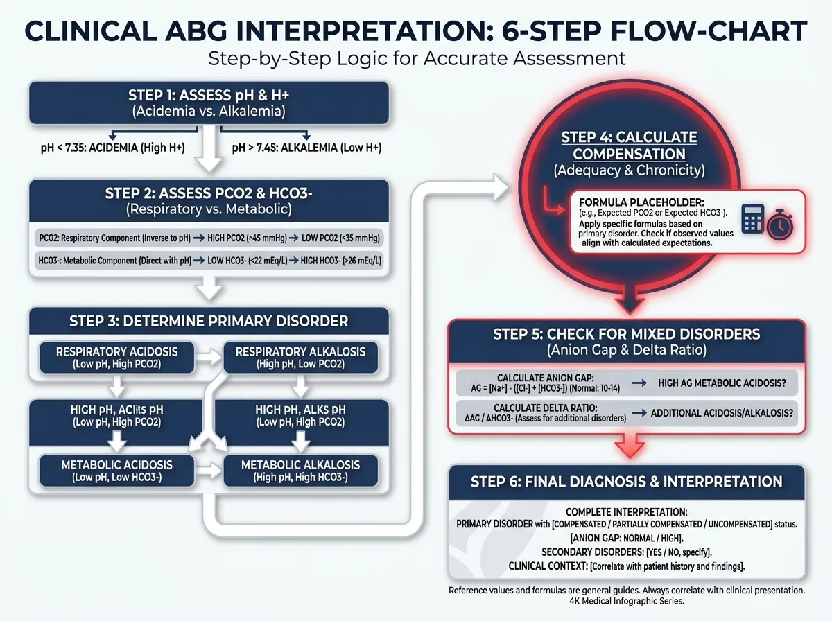
The most reliable way to interpret an ABG — whether you are a nursing student learning ABGs for the first time or an experienced clinician double-checking a bedside calculation — is a systematic six-step approach. Every step builds on the previous, and skipping a step is how mixed disorders get missed.

1
Assess pH Direction. pH < 7.35 = Acidemia. pH > 7.45 = Alkalemia. pH 7.35–7.45 = Normal (may still have a compensated or mixed disorder). This tells you which way the body is tipping.
2
Identify the Primary Disturbance. Look for which parameter — PaCO₂ or HCO₃⁻ — is moving in the same direction as the pH deviation. That is the primary disorder. If PaCO₂ is high and pH is low → Respiratory Acidosis. If HCO₃⁻ is low and pH is low → Metabolic Acidosis.
3
Assess Compensation. Apply the appropriate compensation formula. For metabolic acidosis, use Winter's Formula. For respiratory disorders, use the acute/chronic HCO₃⁻ change rules. If compensation exceeds or falls short of expected, a second primary disorder exists.
4
Calculate Anion Gap (if metabolic acidosis). AG = Na⁺ − (Cl⁻ + HCO₃⁻). Correct for albumin if needed. An elevated AG points to HAGMA (MUDPILES). A normal AG points to NAGMA (hyperchloraemic acidosis). If AG is elevated, calculate the delta-delta ratio to detect concurrent disorders.
5
Assess Oxygenation. Calculate the P/F ratio (PaO₂ ÷ FiO₂) and A-a gradient. An elevated A-a gradient with hypoxaemia suggests V/Q mismatch, shunt, or diffusion defect. A normal A-a gradient with hypoxaemia suggests hypoventilation or low FiO₂.
6
Correlate with Clinical Picture. Numbers never tell the whole story. Check the H-H consistency, review trend (how values have changed over time), and always integrate the ABG with the patient's history, medications, and clinical examination findings.

The ABG Tic-Tac-Toe Method — Explained for Nursing

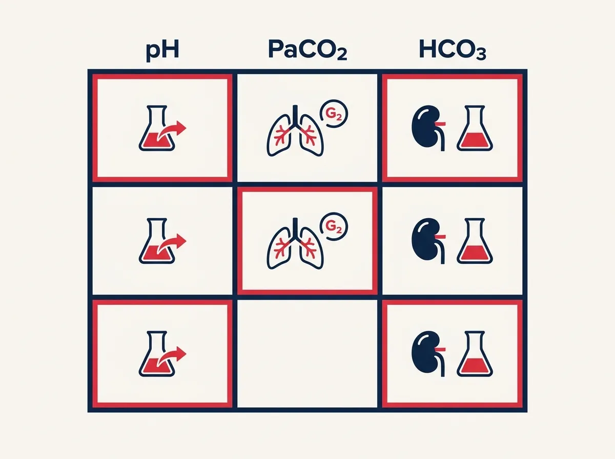
The tic-tac-toe ABG method (also called the ABG grid or 3-box method) is one of the most popular teaching tools in nursing education for quickly identifying primary acid-base disorders. It is especially useful for nursing students learning to calculate ABGs and interpret results at the bedside without needing to memorise complex algorithms. Our ABG calculator includes an interactive tic-tac-toe visual that highlights automatically after each calculation.

The tic-tac-toe grid places pH, PaCO₂, and HCO₃⁻ side by side with arrows indicating their direction relative to normal. The rule is simple: the two boxes that move in the same direction as each other identify the disorder and the compensation. When pH goes acidotic (↓), look to see whether PaCO₂ is high (↑) — if so, it is respiratory acidosis. If HCO₃⁻ is low (↓), it is metabolic acidosis. The third box showing a change represents compensation.

Quick Tic-Tac-Toe Rule: The two boxes that match the pH direction are your disorder. The third box that moves in the opposite direction to the disorder identifies compensation in progress. When all three boxes show abnormal values, always suspect a mixed disorder — this is where a compensation formula is essential to sort it out.

ABG Calculation Practice — 5 Clinical Case Quiz

The best way to master how to calculate ABGs is through regular practice with real clinical scenarios. These five cases cover the most commonly encountered acid-base patterns. Choose your answer, then read the full explanation — each case breaks down the ABG calculation step by step.

pH
PaCO₂
HCO₃⁻
PaO₂
What is the primary acid-base disorder?

Frequently Asked Questions About ABG Calculation

How do you calculate ABGs step by step? +
ABG calculation follows a 6-step systematic approach: (1) Assess pH direction — below 7.35 is acidemia, above 7.45 is alkalemia. (2) Identify the primary disturbance — the parameter moving in the same direction as pH is the primary cause. (3) Assess compensation using Winter's formula for metabolic acidosis or the acute/chronic rules for respiratory disorders. (4) Calculate the anion gap if metabolic acidosis is present. (5) Assess oxygenation via P/F ratio and A-a gradient. (6) Correlate everything with the patient's clinical picture. Enter your values into our ABG calculator above for instant step-by-step analysis.
Is bicarbonate (HCO₃⁻) on an ABG measured or calculated? +
Bicarbonate (HCO₃⁻) is calculated, not directly measured. A blood gas analyser directly measures only three values using electrodes: pH, PaCO₂, and PaO₂. Bicarbonate is then calculated by the machine using the Henderson-Hasselbalch equation: HCO₃⁻ = 0.03 × PaCO₂ × 10^(pH − 6.1). Standard bicarbonate, base excess, and total CO₂ are also all calculated values. This is why our ABG calculator includes a Henderson-Hasselbalch consistency check — to flag when a calculated pH doesn't match the reported values, which can indicate a sampling error.
How to calculate PF ratio from ABG? +
The P/F ratio is calculated by dividing the partial pressure of arterial oxygen (PaO₂ in mmHg, from the ABG) by the fraction of inspired oxygen (FiO₂, expressed as a decimal). P/F ratio = PaO₂ ÷ FiO₂. Example: PaO₂ = 70 mmHg, patient on 40% oxygen (FiO₂ = 0.40) → P/F = 70 ÷ 0.40 = 175. A P/F ratio below 300 meets the Berlin criteria for ARDS; below 200 is severe ARDS. Enter PaO₂ and FiO₂ in our calculator above and the P/F ratio is calculated automatically.
How to calculate ABG compensation? +
Compensation calculation uses the correct formula for each disorder. For metabolic acidosis: apply Winter's Formula — Expected PaCO₂ = 1.5 × HCO₃⁻ + 8 (±2). Compare measured PaCO₂ to this range; a mismatch indicates a second primary disorder. For metabolic alkalosis: Expected PaCO₂ = 0.7 × HCO₃⁻ + 20 (±5). For respiratory acidosis: acute HCO₃⁻ rise = 1 mEq/L per 10 mmHg CO₂ rise; chronic = 3.5 mEq/L. For respiratory alkalosis: acute HCO₃⁻ fall = 2 mEq/L per 10 mmHg CO₂ drop; chronic = 5 mEq/L. If the measured compensation perfectly matches the expected value, it is a simple (single) disorder. If it falls outside this range, a mixed disorder is present.
How to calculate anion gap from ABG? +
The anion gap is calculated using serum electrolytes — not from the ABG directly, though it is used in conjunction with ABG interpretation. The formula is: AG = Na⁺ − (Cl⁻ + HCO₃⁻). The HCO₃⁻ value used is from the ABG report. Normal anion gap is approximately 12 mEq/L (range 8–16). For patients with hypoalbuminaemia, apply the albumin correction: Corrected AG = AG + 2.5 × (4.0 − albumin in g/dL). An elevated corrected AG confirms High Anion Gap Metabolic Acidosis (HAGMA). The MUDPILES mnemonic covers the most common causes.
What is a good ABG value — how do you know if ABG is fully compensated? +
An ABG is fully compensated when the pH has returned to the normal range (7.35–7.45) despite both PaCO₂ and HCO₃⁻ still being abnormal. The key point is that compensation should never normalise the pH perfectly — if it does, suspect a mixed disorder where two opposing primary processes are cancelling each other out. A fully compensated disorder means the compensatory mechanism has been working for enough time (hours for respiratory, days for renal) and has been sufficient to bring pH back into range. The underlying abnormality in PaCO₂ or HCO₃⁻ remains, even when compensated.
How to calculate A-a gradient from ABG? +
The A-a gradient (alveolar-arterial oxygen difference) measures the difference between the calculated alveolar oxygen tension (PAO₂) and the measured arterial oxygen tension (PaO₂). Step 1: Calculate PAO₂ using the alveolar gas equation: PAO₂ = FiO₂ × (Patm − PH₂O) − PaCO₂/RQ = FiO₂ × 713 − PaCO₂/0.8 (at sea level, RQ = 0.8). Step 2: A-a gradient = PAO₂ − PaO₂. The normal A-a gradient is approximately 2.5 + (0.21 × age) mmHg — it widens with age. An elevated A-a gradient indicates that oxygen is being lost between the alveoli and the bloodstream due to V/Q mismatch, intrapulmonary shunt, or diffusion defect.
How to calculate base excess and base deficit from ABG? +
Base excess (BE) is calculated by the blood gas analyser and represents the amount of strong acid needed to titrate one litre of fully oxygenated blood to pH 7.4 at 37°C and PaCO₂ of 40 mmHg. Normal range is −2 to +2 mEq/L. A base excess below −2 (negative BE = base deficit) indicates metabolic acidosis — the metabolic component of the disorder. A base excess above +2 indicates metabolic alkalosis. The magnitude of the deficit tells you approximately how much bicarbonate is needed for correction: HCO₃⁻ deficit (mEq) = Base Deficit × Body Weight (kg) × 0.3. Base excess is preferred in the Copenhagen/Scandinavian approach to ABG interpretation, while the Boston approach uses HCO₃⁻ directly.
How accurate are ABG calculators? +
ABG calculators applying validated formulas — like Winter's formula, the anion gap equation, and the P/F ratio formula — are mathematically exact. They perform the same arithmetic as you would by hand, eliminating calculation errors which are a known source of clinical mistakes. However, the accuracy of the interpretation depends entirely on the accuracy of the input values. ABG samples can be affected by air contamination, delayed processing, incorrect anticoagulant, or arterial vs venous mislabelling. Always verify that the reported values are internally consistent (Henderson-Hasselbalch check), and always interpret in clinical context. This calculator is a tool to aid and teach — not to replace clinical reasoning.
How to calculate PaO₂ from ABG and what does it indicate? +
PaO₂ is directly measured by the blood gas analyser — it is not calculated. It represents the partial pressure of dissolved oxygen in arterial blood. Normal range is 80–100 mmHg on room air (FiO₂21%). PaO₂ below 80 mmHg indicates hypoxaemia. Severity classification: Mild = 60–79 mmHg, Moderate = 40–59 mmHg, Severe = below 40 mmHg. PaO₂ naturally decreases with age: expected PaO₂ ≈ 100 − (0.3 × age in years). PaO₂ must always be interpreted alongside FiO₂ — a PaO₂ of 80 on 100% oxygen is profoundly abnormal, while the same value on room air is normal.

Key Takeaways for ABG Interpretation

pH, PaCO₂, and PaO₂ are directly measured. HCO₃⁻, base excess, and standard bicarbonate are all calculated values.
The primary disorder is identified by the parameter moving in the same direction as pH — respiratory (CO₂) or metabolic (HCO₃⁻).
Winter's formula (Expected PaCO₂ = 1.5 × HCO₃⁻ + 8 ±2) is the single most clinically important compensation formula to memorise.
P/F ratio below 300 = ARDS criteria. Below 200 = Severe ARDS. Always calculate when both PaO₂ and FiO₂ are known.
Always apply the albumin correction to the anion gap — hypoalbuminaemia reduces the normal AG, potentially masking a high-AG acidosis.
🩺
Dr. Heart (Health Observer)
Cardiovascular & Critical Care Health Specialist

All formulas in this ABG calculator are referenced against peer-reviewed clinical literature: Winter's formula (Winters et al., 1967), the Boston compensation rules (Schwartz et al., Brenner et al.), anion gap equations (Emmett & Narins, 1977), the P/F ratio (Horowitz et al., 1974), and the A-a gradient equation. Practice cases are based on common clinical presentations reviewed against UCSF Hospital Handbook and ATS guidelines. Content is written for educational accuracy and clinical usefulness.