What Is the Wells Score for PE and DVT?
The Wells Score (Wells Criteria) is the most widely validated clinical prediction algorithm in emergency medicine and cardiology for estimating the pre-test probability of venous thromboembolism (VTE). Developed by Dr. Philip S. Wells and colleagues at the University of Ottawa, the model was established in two clinical formulations: the Wells Criteria for Pulmonary Embolism (PE) (Wells et al. 2000, 2001) and the Wells Criteria for Deep Vein Thrombosis (DVT) (Wells et al. 1997, 2003).
The foundational clinical purpose of the Wells scoring system is to safely guide diagnostic testing in patients presenting with acute pleuritic chest pain, dyspnea, or unilateral leg swelling. By integrating objective signs (such as tachycardia, immobilization, or asymmetric calf enlargement) with subjective clinical gestalt (whether an alternative diagnosis is equally or less likely), clinicians determine whether a patient can be safely evaluated with a non-invasive high-sensitivity D-dimer blood test or requires immediate diagnostic imaging via CT Pulmonary Angiography (CTPA) or Compression Duplex Ultrasonography (CUS).
How Is the Wells Score Calculated?
The Wells score assigns differential integer and half-integer mathematical weightings based on the relative odds ratios determined in prospective observational derivation trials. The tool supports two distinct diagnostic workflows:
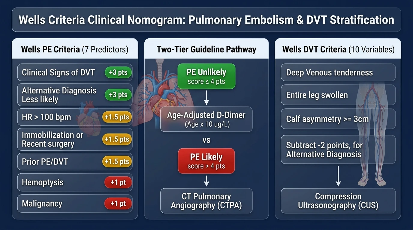
1. Wells Criteria for Pulmonary Embolism (7 Clinical Variables)
The PE model sums points across 7 clinical determinants, yielding a cumulative score ranging from 0 to 12.5 points:
| Clinical Predictor | Clinical Definition & Diagnostic Basis | Points |
|---|---|---|
| Clinical signs & symptoms of DVT | Minimum of objective swelling of the leg and deep venous palpation pain. | +3.0 pts |
| Alternative diagnosis less likely than PE | Physician gestalt that PE is the most likely or equally likely etiology for symptoms. | +3.0 pts |
| Heart rate > 100 beats / minute | Resting tachycardia reflecting sympathetic activation or right ventricular strain. | +1.5 pts |
| Immobilization (≥ 3 days) or surgery (< 4 weeks) | Consecutive bed rest for at least 3 days or operative intervention under general anesthesia. | +1.5 pts |
| Previous documented DVT or PE | Prior objectively confirmed history of venous thromboembolism. | +1.5 pts |
| Hemoptysis | Coughing up frank blood or blood-tinged sputum indicating pulmonary infarction. | +1.0 pt |
| Malignancy | Active cancer treatment within past 6 months or receiving palliative oncological care. | +1.0 pt |
2. Wells Criteria for Deep Vein Thrombosis (10 Clinical Variables)
The DVT model evaluates 10 clinical signs, notably including a 2-point penalty deduction when an alternative diagnosis is at least as likely as DVT. The total score ranges from -2 to 9 points:
| DVT Clinical Predictor | Diagnostic Criteria & Measurement | Points |
|---|---|---|
| Active cancer | Treatment ongoing, within 6 months, or palliative care. | +1.0 pt |
| Paralysis, paresis, or recent plaster cast | Neurological deficit or orthopedic immobilization of lower extremity. | +1.0 pt |
| Bedridden > 3 days or major surgery (< 12 weeks) | Recent major abdominal, pelvic, or orthopedic surgery requiring regional/general anesthesia. | +1.0 pt |
| Localized tenderness | Tenderness along anatomical distribution of deep venous system (femoral/popliteal). | +1.0 pt |
| Entire leg swollen | Unilateral diffuse swelling from thigh to foot. | +1.0 pt |
| Calf swelling ≥ 3 cm asymmetric | Measured circumference 10 cm below tibial tuberosity compared to asymptomatic calf. | +1.0 pt |
| Pitting edema confined to symptomatic leg | Unilateral pitting edema without systemic bilateral congestive heart failure. | +1.0 pt |
| Collateral superficial veins (non-varicose) | Visible distended superficial veins over the tibia or thigh. | +1.0 pt |
| Previously documented DVT | Prior ultrasound or venogram documenting deep venous thrombosis. | +1.0 pt |
| Alternative diagnosis as likely as DVT | Identifiable alternative etiology (Baker's cyst, cellulitis, muscle tear, hematoma). | -2.0 pts |
Two-Tier vs Three-Tier Risk Stratification: Which Model to Use?
Historically, Dr. Wells proposed a Three-Tier System dividing patients into Low, Moderate, and High clinical probability. However, contemporary international guidelines from the American College of Emergency Physicians (ACEP), European Society of Cardiology (ESC 2019), and British Thoracic Society (BTS) endorse the Two-Tier Model ("PE Unlikely" vs "PE Likely") as the superior standard of care.
| Model System | Point Cutoff | Clinical Category | Observed PE Prevalence | Guideline Diagnostic Strategy |
|---|---|---|---|---|
| Two-Tier Model (Guideline Standard) |
≤ 4.0 Points | PE Unlikely | ~8.0% – 12.0% | High-sensitivity D-Dimer (Rule out if negative; CTPA only if positive) |
| > 4.0 Points | PE Likely | ~28.0% – 37.0% | Immediate CT Pulmonary Angiography (Do not wait for D-dimer) | |
| Three-Tier Model (Traditional) |
< 2.0 Points | Low Probability | ~3.6% (1.5% – 5.0%) | High-sensitivity D-Dimer assay |
| 2.0 – 6.0 Points | Moderate Probability | ~16.2% (12.0% – 28.0%) | High-sensitivity D-Dimer or direct CTPA based on clinical context | |
| > 6.0 Points | High Probability | ~40.6% – 65.0% | Immediate CTPA; consider empiric anticoagulation if delay expected |
Why the Two-Tier Model Is Guideline-Preferred
Large prospective multi-center trials (including the Christopher Study Group in JAMA 2006) demonstrated that binary categorization into "PE Unlikely" (≤ 4 points) combined with a high-sensitivity D-dimer safely ruled out PE in over 30% of emergency department presentations with an exceptionally low 3-month thromboembolic failure rate of only 0.5% (95% CI: 0.2%–1.1%). This eliminated ambiguous intermediate-probability categories that frequently led to unnecessary, low-yield CT radiation.
The Age-Adjusted D-Dimer Formula: Safely Avoiding Unnecessary CT Scans
Plasma D-dimer levels naturally increase with advancing chronological age due to subclinical endothelial activation, chronic low-grade vascular inflammation, and progressive decline in renal clearance (frequently assessed with the Creatinine Clearance Calculator or GFR Calculator). Consequently, using a rigid unadjusted cutoff of 500 µg/L in elderly patients yields a specificity of less than 10% in patients over 80 years of age, causing almost all elderly patients to undergo CTPA.
To overcome this limitation, the ADJUST-PE Study (Righini et al., JAMA 2014) validated the Age-Adjusted D-Dimer Rule for patients older than 50 years with an "Unlikely" pre-test probability:
(Applicable only for patients aged > 50 years; patients ≤ 50 maintain standard 500 µg/L cutoff)
- Patient Aged 50 or Younger: Normal cutoff remains 500 µg/L.
- Patient Aged 65 Years: Age-adjusted cutoff is 650 µg/L (65 × 10).
- Patient Aged 75 Years: Age-adjusted cutoff is 750 µg/L (75 × 10).
- Patient Aged 85 Years: Age-adjusted cutoff is 850 µg/L (85 × 10).
Implementing the age-adjusted cutoff increases the diagnostic efficiency of D-dimer in patients > 75 years four-fold (from 6.4% to 29.7%) without increasing false-negative rates, preserving a post-test 3-month thromboembolic risk well below 1%.
Wells Score vs Geneva Score vs PERC Rule: Emergency Decision Framework
Emergency clinicians often choose between three complementary clinical decision rules when evaluating suspected pulmonary embolism:
| Decision Algorithm | Clinical Focus & Target Group | Key Strength | Notable Limitation |
|---|---|---|---|
| Wells Criteria (PE) | General suspected PE (ED & inpatient) | Highest clinical validation and guideline integration globally; dual 2-tier/3-tier options | Includes subjective clinician gestalt ("alternative diagnosis less likely", 3 pts) |
| Revised Geneva Score | Suspected PE (inpatient & outpatient) | 100% objective variables (no subjective clinician gestalt required) | Slightly lower specificity compared to Wells in younger emergency cohorts |
| PERC Rule | Very low-risk suspected PE only | Can rule out PE without a blood draw if all 8 criteria are negative | Must NEVER be applied to intermediate or high-risk patients; missing 1 criteria invalidates rule |
The Stepped Emergency Triage Hierarchy: Clinicians first evaluate low-acuity chest pain patients. If the patient has a very low gestalt probability and meets all 8 PERC criteria, workup stops without testing. If PERC is positive or gestalt is moderate/high, clinicians apply the Wells Score to categorize pre-test probability and govern D-dimer versus CTPA imaging.
Diagnostic Imaging Protocols: CTPA, V/Q Scans & Compression Ultrasound
When pre-test probability is likely (Wells > 4.0) or D-dimer is positive, definitive imaging confirms or excludes clot location:
- CT Pulmonary Angiography (CTPA): The gold-standard imaging modality (sensitivity 83%–100%, specificity 89%–97%). Directly visualizes intraluminal filling defects in main, lobar, segmental, and subsegmental pulmonary arteries. Interventionalists track Mean Arterial Pressure (MAP) and right ventricular strain on echocardiography when massive clot burden is detected.
- Ventilation / Perfusion (V/Q) Scintigraphy: First-line alternative when iodinated contrast is contraindicated (severe renal impairment, severe contrast anaphylaxis, or uninstrumented pregnancy). A high-probability V/Q scan (mismatched segmental perfusion defects) confirms PE; a normal scan completely excludes it.
- Compression Duplex Ultrasonography (CUS): The primary diagnostic test for suspected Deep Vein Thrombosis. Visualizes loss of deep vein compressibility in the common femoral, femoral, and popliteal veins. In patients with suspected PE who have obvious DVT signs, a positive proximal CUS provides sufficient justification for therapeutic anticoagulation, potentially sparing a sick patient a trip to the CT scanner.
Clinical Limitations, Hemodynamic Shock & Massive PE
While the Wells score provides robust diagnostic discrimination, clinicians must be alert to critical clinical scenarios where standard scoring pathways do not apply:
⚠️ Massive PE / Hemodynamic Collapse Alert
The Wells score is validated for hemodynamically stable patients. If a patient presents with sustained hypotension (systolic blood pressure < 90 mmHg for > 15 minutes), cardiogenic shock, or cardiac arrest, the patient has High-Risk (Massive) Pulmonary Embolism. Do NOT waste time calculating Wells scores or ordering D-dimer assays. Immediately initiate resuscitative protocols, bedside echocardiography (McConnell's sign, RV dilation), unfractionated heparin, and emergent CTPA or systemic thrombolytic therapy (alteplase / tPA).
- Subjectivity of Criterion #2: Awarding 3.0 points for "PE is #1 diagnosis or equally likely" relies on clinical experience. Inexperienced examiners may underestimate this item in patients with subtle atypical presentations.
- Subsegmental PE Controversies: Modern high-resolution 128-slice CT scanners frequently detect isolated subsegmental pulmonary emboli (SSPE) of uncertain clinical significance. Management requires individualized multidisciplinary discussion regarding anticoagulation versus surveillance.
- Concomitant Cardiovascular Syndromes: Patients presenting with acute chest pain must be concurrently screened for acute coronary syndromes using validated tools such as the HEART Score for Chest Pain, TIMI Risk Score, and GRACE Score Calculator to avoid misattributing myocardial ischemia to pleuritic pain.
Real-World Clinical Case Studies
Case 1: 67-Year-Old Female with Post-Operative Tachycardia
PE Unlikely (D-Dimer Protocol)Clinical Scenario: A 67-year-old female presents to the urgent assessment unit 10 days following elective total hip arthroplasty. She reports mild shortness of breath upon exertion. Vital signs: pulse 104 bpm, blood pressure 130/78 mmHg, SpO2 97% on room air. Her surgical leg has expected healing incisions with mild symmetrical postoperative edema without localized deep vein tenderness. She has no hemoptysis or active cancer.
Clinical Pathway & Outcome: A score of 3.0 places her in the "PE Unlikely" tier (≤ 4.0 points). Under the age-adjusted protocol, her decision threshold is 670 µg/L (67 × 10). Her high-sensitivity D-dimer returns at 540 µg/L (which would have been flagged as elevated by a rigid 500 cutoff). Because 540 µg/L is below her age-adjusted threshold of 670 µg/L, pulmonary embolism is safely ruled out, avoiding contrast nephropathy and CT radiation.
Case 2: 52-Year-Old Male with Painful Unilateral Leg Swelling
DVT Likely (Ultrasound Protocol)Clinical Scenario: A 52-year-old male with active colon adenocarcinoma (undergoing adjuvant FOLFOX chemotherapy) presents with a 3-day history of worsening left calf pain. Physical examination demonstrates marked localized tenderness along the popliteal vein, pitting edema confined to the left leg, and a left calf circumference of 39 cm compared to 34 cm on the right (+5 cm asymmetry). There are no symptoms of superficial cellulitis or trauma.
Clinical Pathway & Outcome: A Wells DVT score of 4.0 classifies the patient as "DVT Likely" (≥ 2 points) with an estimated disease prevalence > 40%. In accordance with guideline pathways, D-dimer testing is skipped. The patient undergoes urgent lower extremity Compression Duplex Ultrasonography (CUS), which confirms an acute occlusive thrombus in the left popliteal and posterior tibial veins. Therapeutic low-molecular-weight heparin is promptly initiated.
Frequently Asked Questions About the Wells Score
What does the Wells Score calculate?
The Wells Score calculates the pre-test clinical probability of venous thromboembolism (VTE) across two distinct validated models: Wells Criteria for Pulmonary Embolism (PE) and Wells Criteria for Deep Vein Thrombosis (DVT). It guides clinicians on whether to order a non-invasive D-dimer test or proceed directly to definitive diagnostic imaging.
What is the difference between the 2-tier and 3-tier Wells PE models?
The traditional 3-tier model categorizes PE probability into Low (< 2.0 pts), Moderate (2.0–6.0 pts), and High (> 6.0 pts). The guideline-preferred 2-tier model simplifies stratification into PE Unlikely (≤ 4.0 pts) and PE Likely (> 4.0 pts). Clinical trials demonstrate that the 2-tier system provides superior diagnostic efficiency without increasing missed venous thromboembolism events.
How is the Age-Adjusted D-Dimer threshold calculated?
For patients older than 50 years with an 'Unlikely' pre-test probability, the age-adjusted D-dimer cutoff is calculated as: Age (years) × 10 µg/L (or Age × 0.01 mg/L in FEU units). For example, a 70-year-old patient has an age-adjusted cutoff of 700 µg/L rather than the standard 500 µg/L threshold, safely reducing unnecessary CTPA imaging by over 20% in elderly populations.
When should a D-Dimer test be avoided in suspected PE?
A D-dimer test should be avoided when a patient is categorized as 'PE Likely' (Wells score > 4.0) or 'High Probability' (Wells score > 6.0). In high-risk tiers, a negative D-dimer is insufficient to safely rule out PE due to an unacceptable false-negative rate (~15%); these patients should proceed directly to CT Pulmonary Angiography (CTPA) without testing delay.
Why does the Wells DVT score subtract 2 points for an alternative diagnosis?
In the Wells DVT model, having an alternative diagnosis at least as likely as DVT (such as Baker's cyst, cellulitis, superficial thrombophlebitis, or calf muscle tear) subtracts 2 points. This penalty accounts for common clinical mimics and significantly lowers false-positive pre-test probabilities, preventing unwarranted compression ultrasonography.
How does the Wells PE Score differ from the PERC Rule?
The PERC Rule (Pulmonary Embolism Rule-out Criteria) consists of 8 high-sensitivity criteria designed solely for very low-risk emergency patients. If a patient is deemed clinically low risk by gestalt and meets ALL 8 PERC criteria, PE can be ruled out without even drawing a D-dimer. In contrast, the Wells Score is a comprehensive quantitative pre-test model used to categorize intermediate and higher-risk clinical presentations.
What is the diagnostic imaging of choice for PE and DVT?
For suspected Pulmonary Embolism with a likely pre-test probability or positive D-dimer, multidetector CT Pulmonary Angiography (CTPA) is the gold standard imaging modality. For suspected Deep Vein Thrombosis, whole-leg or proximal Compression Duplex Ultrasonography (CUS) is the definitive initial diagnostic test.
Peer-Reviewed Medical References & Clinical Guidelines
- Wells PS, Anderson DR, Rodger M, et al. Derivation of a simple clinical model to categorize patients probability of pulmonary embolism: increasing the models utility with the SimpliRED D-dimer. Thromb Haemost. 2000;83(3):416-420.
- Wells PS, Anderson DR, Rodger M, et al. Excluding pulmonary embolism at the bedside without diagnostic imaging: management of patients with suspected pulmonary embolism presenting to the emergency department by using a simple clinical model and d-dimer. Ann Intern Med. 2001;135(2):98-107. doi:10.7326/0003-4819-135-2-200107170-00010
- Wells PS, Anderson DR, Bormanis J, et al. Value of assessment of pretest probability of deep-vein thrombosis in clinical management. Lancet. 1997;350(9094):1795-1798. doi:10.1016/S0140-6736(97)08460-2
- Righini M, Van Es J, Den Exter PL, et al. Age-adjusted D-dimer cutoff levels to rule out pulmonary embolism: the ADJUST-PE study. JAMA. 2014;311(11):1117-1124. doi:10.1001/jama.2014.2135
- Konstantinides SV, Meyer G, Becattini C, et al. 2019 ESC Guidelines for the diagnosis and management of acute pulmonary embolism developed in collaboration with the European Respiratory Society (ERS). Eur Heart J. 2020;41(4):543-603. doi:10.1093/eurheartj/ehz405
- van der Hulle T, Cheung WY, Kooij S, et al. Simplified diagnostic management of suspected pulmonary embolism (the YEARS study): a prospective, multicentre, cohort study. Lancet. 2017;390(10091):289-297. doi:10.1016/S0140-6736(17)30885-1