Physiological Foundations of Fractional Excretion of Urea
The Fractional Excretion of Urea (FEUrea)—frequently referred to in United States clinical literature as the Fractional Excretion of Urea Nitrogen (FEUN)—measures the percentage of filtered urea that escapes renal tubular reabsorption and is excreted in the urine. By indexing urea clearance directly to creatinine clearance, the calculation eliminates the confounding effect of urine concentration or dilution in spot urine samples.
In human renal physiology, urea and creatinine exhibit radically distinct tubular kinetics:
- Serum Creatinine: Creatinine is freely filtered across the glomerular filtration barrier and undergoes virtually zero tubular reabsorption (with only 10% active secretion by proximal organic cation transporters). Consequently, creatinine clearance serves as a reliable denominator mirroring true glomerular filtration rate (GFR).
- Urea & Urea Nitrogen: Urea is also freely filtered at the glomerulus. However, approximately 40% to 50% of filtered urea is passively reabsorbed in the proximal convoluted tubule via paracellular solvent drag coupled to sodium and water reabsorption. Additional regulated urea reabsorption occurs in the inner medullary collecting duct via vasopressin-sensitive urea transporters (UT-A1 and UT-A3).
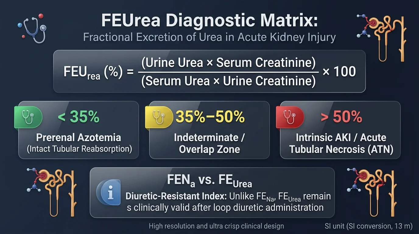
When renal hypoperfusion occurs (prerenal azotemia), the baroreceptor reflex and renin-angiotensin-aldosterone system (RAAS) drive avid proximal tubular sodium and water reabsorption. As fluid reabsorption slows tubular flow velocity, urea reabsorption accelerates dramatically, dropping urinary urea excretion and driving the FEUrea below 35%. Conversely, when ischemic or nephrotoxic injury destroys tubular brush border epithelial cells (Acute Tubular Necrosis / ATN), tubular reabsorption fails, and filtered urea washes into the collecting duct, driving FEUrea above 50%.
Assess full renal trajectories by pairing FEUrea with the FENa Calculator, evaluate prerenal filtration disproportions via the BUN:Creatinine Ratio Calculator, and monitor hourly catheter output with the Urine Output Calculator.
Mathematical Formula & Clearance Derivation
Fractional excretion represents the mathematical ratio of urea clearance ($C_{\text{urea}}$) to creatinine clearance ($C_{\text{Cr}}$), multiplied by 100 to yield a percentage:
Pairs serum Blood Urea Nitrogen (BUN) with spot Urine Urea Nitrogen (UUN) in mg/dL.
Pairs total serum urea with total urine urea in mmol/L, and creatinine in µmol/L.
The Diuretic Advantage: Why FEUrea Bypasses FENa Failure
In hospitalized patients evaluated for acute kidney injury, the Fractional Excretion of Sodium (FENa) is frequently ordered first. However, in modern clinical medicine, more than 60% of patients with acute azotemia have recently received loop diuretics (such as furosemide, bumetanide, or torsemide) or thiazide diuretics for congestive heart failure, volume overload, or hypertension.
This creates a diagnostic pitfall:
- How Loop Diuretics Invalidate FENa: Loop diuretics selectively inhibit the $\text{Na}^+\text{-K}^+\text{-2Cl}^-$ symporter in the thick ascending limb of Henle's loop. This blocks approximately 25% of filtered sodium reabsorption, triggering profuse natriuresis. Consequently, a patient with severe prerenal hypoperfusion who receives 40 mg of IV furosemide will excrete large amounts of urinary sodium, driving their FENa from $0.4\%$ up to $2.5\%\text{--}4.0\%$. A clinician relying on FENa would mistakenly diagnose irreversible Acute Tubular Necrosis (ATN) and withhold lifesaving volume restoration.
- Why FEUrea Remains Intact: Urea reabsorption occurs primarily in the proximal convoluted tubule (S1–S3 segments) through passive paracellular movement and solvent drag. Loop diuretics act downstream in the loop of Henle and do not block proximal tubular urea reabsorption. Therefore, in prerenal states, proximal tubular avidity for urea continues unabated, keeping the FEUrea reliably below 35% despite massive furosemide-induced natriuresis.
Numerous prospective validation trials (including landmark studies by Carvounis et al. and Pépin et al.) demonstrated that in diuretic-treated patients with acute kidney injury, FEUrea maintains a diagnostic sensitivity of 89% and specificity of 83% for prerenal azotemia, compared to FENa, whose diagnostic accuracy drops to near random chance.
Step-by-Step Clinical Protocol for FEUrea Calculation
-
1
Obtain Simultaneously Paired Blood and Urine Samples
Ensure the serum chemistry panel (BUN and creatinine) and the spot urine specimen (UUN and urine creatinine) are collected at the same time. Comparing morning serum labs with evening post-resuscitation urine invalidates the clearance ratio.
-
2
Confirm Laboratory Analyte Alignment
Verify whether the laboratory reports Blood Urea Nitrogen (BUN) in mg/dL or total Urea in mmol/L. The spot urine test must match the same analyte basis.
-
3
Check Medication Administration History
Document the timing of loop diuretics (furosemide, bumetanide, torsemide) and osmotic agents (mannitol). If diuretics were administered within the preceding 24 hours, document FEUrea as the primary diagnostic index over FENa.
-
4
Execute Fractional Excretion Arithmetic
Multiply urine urea by serum creatinine, divide by serum urea multiplied by urine creatinine, and multiply by 100.
-
5
Classify into Validated Diagnostic Tiers
Categorize the percentage into Prerenal (< 35%), Intermediate Overlap (35%–50%), or Intrinsic Tubular Injury (> 50%).
-
6
Correlate with Urine Microscopy & Hemodynamics
Examine urine sediment for muddy brown granular casts (ATN) versus hyaline casts (prerenal), monitor mean arterial pressure using the Mean Arterial Pressure Calculator, and evaluate glomerular filtration with the GFR Calculator.
Clinical Case Scenarios: Real-World Diagnostic Utility
Case Scenario 1: Cardiorenal Syndrome on IV Loop Diuretics (Prerenal Azotemia)
Patient Presentation: A 67-year-old male with ischemic cardiomyopathy (LVEF 28%) is hospitalized for acute decompensated heart failure with profound lower extremity edema and orthopnea. He receives continuous intravenous furosemide at 10 mg/hr. On hospital day 3, his urine output declines to 25 mL/hr, and his serum creatinine rises acutely from 1.1 to 2.3 mg/dL.
Clinical Synthesis & Management: While the FENa of 2.13% misleadingly points to acute tubular necrosis, the FEUrea of 24.3% reveals preserved proximal tubular urea reabsorption. Active loop diuretic therapy was washing sodium into the urine, invalidating FENa. The patient has type 1 cardiorenal syndrome with renal hypoperfusion driven by poor forward cardiac output and high venous congestion. Rather than stopping diuretics and causing asphyxiating pulmonary edema, the clinical team optimizes cardiac inotropy with low-dose dobutamine, monitors perfusion pressure via the Mean Arterial Pressure Calculator, and tracks volume status with the Urine Output Calculator, leading to creatinine stabilization.
Case Scenario 2: Aminoglycoside Nephrotoxicity & Septic Acute Tubular Necrosis
Patient Presentation: A 54-year-old female in the intensive care unit is mechanically ventilated for severe Klebsiella pneumoniae urosepsis. She has been treated with IV meropenem and high-dose IV gentamicin. On ICU day 5, despite adequate blood pressure resuscitation on norepinephrine, her urine output drops to 15 mL/hr and serum creatinine climbs from 0.9 to 3.4 mg/dL.
Clinical Synthesis & Management: An FEUrea of 57.2% demonstrates failure of proximal tubular epithelial reabsorption. Gentamicin accumulates in proximal tubular lysosomes, causing brush border membrane rupture, mitochondrial swelling, and extensive acute tubular necrosis. Because tubular cells are necrotic, filtered urea is unable to be reabsorbed passively and washes out into the urine. Recognizing true intrinsic ATN, the critical care team immediately discontinues gentamicin, avoids excessive fluid overload, adjusts renal antibiotic dosing using the GFR Calculator, and prepares for possible renal replacement therapy.
Clinical Reference Tables: Diagnostic Cutoffs & Confounder Matrix
The following matrices outline the clinical interpretation of FEUrea values alongside a head-to-head comparison with FENa:
| Diagnostic Clinical Metric | FENa (Fractional Excretion of Sodium) | FEUrea (Fractional Excretion of Urea) | Preferred Clinical Setting |
|---|---|---|---|
| Prerenal Azotemia Cutoff | < 1.0% (Avid sodium reabsorption) | < 35.0% (Avid proximal urea reabsorption) | FEUrea is preferred if diuretics given in past 24 hrs. |
| Intrinsic ATN Cutoff | > 2.0% (Tubular sodium wasting) | > 50.0% (Tubular urea reabsorptive failure) | Either index valid in diuretic-naïve patients. |
| Intermediate Overlap Zone | 1.0% to 2.0% | 35.0% to 50.0% | Requires urine sediment microscopy and clinical correlation. |
| Confounded by Loop Diuretics? | YES — Highly Conflicted: Diuretics cause false-positive elevations (> 2%). | NO — Diuretic-Resistant: Reabsorption occurs in proximal tubule, upstream of Henle's loop. | FEUrea is the diagnostic standard of choice when furosemide or bumetanide is active. |
| Confounded by Osmotic Diuresis? | Yes (Mannitol, severe glucosuria) | Yes (Urea washout decreases reabsorption) | Both indices confounded; evaluate clinical volume directly. |
Diagnostic stratification based on the calculated FEUrea percentage:
| FEUrea Value | Classification | Underlying Nephron Pathophysiology | Recommended Clinical Next Steps |
|---|---|---|---|
| < 35% | Prerenal Azotemia | Intact tubular function; high proximal solvent drag and ADH-mediated medullary urea reabsorption driven by volume depletion. | Assess fluid responsiveness; administer cautious crystalloid bolus; optimize cardiac output in cardiorenal syndrome. |
| 35% to 50% | Intermediate / Overlap Zone | Indeterminate; mixed prerenal and acute tubular injury, baseline CKD, resolving ATN, or mild thiazide diuretic effect. | Review urine microscopy (granular vs. hyaline casts); monitor serum creatinine slope; avoid nephrotoxins. |
| > 50% | Intrinsic AKI / ATN | Loss of proximal tubular brush border integrity; damaged tubular cells cannot reabsorb urea back into peritubular capillaries. | Discontinue nephrotoxic agents (NSAIDs, aminoglycosides, contrast); manage volume overload; anticipate dialysis need. |
Physiological Confounders & Diagnostic Pitfalls
Although FEUrea provides superior diagnostic accuracy in diuretic-exposed patients, clinicians must recognize situations where urea excretion kinetics are altered independently of tubular injury:
- Low-Protein Diet & Hepatic Cirrhosis (Pseudo-Low FEUrea): Malnourished patients, alcoholics, and patients with advanced liver failure generate substantially less urea due to urea cycle impairment. Diminished filtered urea loads can result in an FEUrea below 35% even in the presence of established acute tubular necrosis.
- Advanced Baseline Chronic Kidney Disease (CKD): In patients with severe baseline CKD (eGFR < 30 mL/min/1.73m²), surviving nephrons undergo compensatory hyperfiltration and osmotic solute diuresis, reducing baseline urea reabsorption. In this population, prerenal hypoperfusion may produce an FEUrea in the 35%–50% overlap range rather than < 35%. Verify baseline filtration using the GFR Calculator.
- Septic AKI (Mixed Hemodynamics): Sepsis causes complex intrarenal shunting, efferent arteriolar vasodilation, and microvascular hypoperfusion without overt tubular necrosis. In septic AKI, FEUrea values frequently fall into the 35%–50% indeterminate zone.
- Osmotic Diuresis (Hyperglycemia & Mannitol): Marked glucosuria (blood glucose > 300 mg/dL) or IV mannitol expands tubular fluid volume and accelerates luminal flow, flushing urea into the urine and falsely driving FEUrea above 50%.
- Acetazolamide & SGLT2 Inhibitors: Drugs that directly inhibit proximal tubular sodium-coupled transport can reduce proximal urea reabsorption, causing a modest increase in baseline FEUrea.
- The Diuretic-Resistant Standard: When loop diuretics (furosemide, bumetanide, torsemide) invalidate FENa by inducing massive natriuresis, FEUrea is the validated clinical index of choice for distinguishing prerenal azotemia from ATN.
- Prerenal Cutoff (< 35%): An FEUrea below 35% indicates intact proximal tubular urea reabsorption driven by effective circulating volume depletion or cardiac hypoperfusion.
- Intrinsic Cutoff (> 50%): An FEUrea exceeding 50% indicates loss of tubular epithelial reabsorptive capacity, strongly pointing toward Acute Tubular Necrosis (ATN).
- Beware the 35%–50% Overlap Zone: An intermediate FEUrea requires clinical synthesis with urine microscopy, urinalysis, serum creatinine trajectory, and volume status.
- Strict Pairing Requirement: Serum and spot urine samples must be drawn simultaneously, and matching analytes (BUN with UUN, or total urea with total urea) must be utilized.
FEUrea Calculator Frequently Asked Questions
How do you calculate the Fractional Excretion of Urea (FEUrea)?
FEUrea is calculated from paired serum and spot urine samples using the formula: FEUrea (%) = [(Urine Urea × Serum Creatinine) ÷ (Serum Urea × Urine Creatinine)] × 100. In conventional U.S. units, Urine Urea Nitrogen (UUN, mg/dL) and Blood Urea Nitrogen (BUN, mg/dL) are paired with serum and urine creatinine (mg/dL). In SI units, urine and serum urea are measured in mmol/L and paired with creatinine in µmol/L.
Why is FEUrea superior to FENa in patients receiving loop diuretics?
Loop diuretics (such as furosemide, bumetanide, or torsemide) inhibit the Na-K-2Cl cotransporter in the thick ascending limb of Henle's loop, causing massive urinary sodium excretion even when severe prerenal hypoperfusion exists. This artificially elevates the Fractional Excretion of Sodium (FENa) above 1% or 2%, falsely mimicking acute tubular necrosis (ATN). In contrast, urea is primarily reabsorbed passively in the proximal convoluted tubule via solvent drag and sodium-coupled water transport, and in the inner medullary collecting duct via UT-A1/UT-A3 transporters. Because loop diuretics do not directly inhibit proximal tubular urea reabsorption, FEUrea remains below 35% in prerenal azotemia despite active diuretic therapy.
What does an FEUrea below 35% indicate in acute kidney injury?
An FEUrea below 35% indicates high renal tubular avidity for urea, typical of prerenal azotemia (renal hypoperfusion). When effective circulating volume falls (due to dehydration, hemorrhage, congestive heart failure, or cirrhosis), proximal tubular sodium and water reabsorption surges. This slows tubular fluid flow and allows extensive passive reabsorption of filtered urea back into peritubular capillaries, resulting in low fractional urea excretion into the urine.
What does an FEUrea above 50% indicate?
An FEUrea above 50% reflects impaired tubular urea reabsorption, characteristic of intrinsic renal acute kidney injury, most commonly ischemic or nephrotoxic Acute Tubular Necrosis (ATN). Widespread necrosis or shedding of proximal tubular brush border epithelial cells prevents the kidney from reabsorbing urea, causing filtered urea to pass directly into the urine.
How do you interpret an intermediate FEUrea between 35% and 50%?
An FEUrea between 35% and 50% represents a non-diagnostic overlap zone. This intermediate pattern commonly occurs in evolving or resolving AKI, mixed prerenal and intrinsic pathologies (e.g., cardiorenal syndrome transitioning into ATN), patients with underlying chronic kidney disease (CKD), or patients on thiazide diuretics. Clinicians should correlate this result with urine microscopy, urinalysis, serum creatinine trajectory, and volume status rather than relying solely on the cutoff.
Can total serum urea in mmol/L be mixed with Blood Urea Nitrogen (BUN) in mg/dL?
No. Total urea measures the molecular weight of the whole urea molecule (60.06 g/mol), whereas Blood Urea Nitrogen (BUN) measures only the mass of the two nitrogen atoms in urea (28.01 g/mol). Serum and urine must both use total urea (SI mmol/L) or both use urea nitrogen (US mg/dL). Mixing total urea with urea nitrogen produces an invalid ratio distorted by a factor of 2.14.
What clinical conditions can cause a falsely high or low FEUrea?
A falsely low FEUrea (< 35%) can occur in severe malnutrition or low-protein diets due to diminished urea production, or in acute glomerulonephritis where tubular integrity is preserved despite profound filtration decline. A falsely elevated FEUrea (> 50%) can occur during osmotic diuresis (severe hyperglycemia, mannitol infusion, or urea washout), advanced baseline CKD with fixed solute excretion, or prolonged acetazolamide (proximal tubular diuretic) therapy.
Evidence & Clinical Literature
- Carvounis CP, Nisar S, Guro-Razuman S. Significance of the fractional excretion of urea in the differential diagnosis of acute renal failure. Kidney International. 2002;62(6):2223-2229.
- Pépin MN, Bouchard J, Legault L, Ethier J. Diagnostic performance of fractional excretion of urea and fractional excretion of sodium in the evaluations of patients with acute kidney injury with or without diuretics. American Journal of Kidney Diseases. 2007;50(4):566-573.
- Darmon M, Vincent F, Dellamonica J, et al. Diagnostic performance of fractional excretion of urea in the evaluation of critically ill patients with acute kidney injury: a multicenter cohort study. Critical Care. 2011;15(4):R178.
- Diskin CJ, Stokes TJ, Dansby LM, et al. The fractional excretion of urea: a critical review of its utility in the evaluation of acute kidney injury. Renal Failure. 2010;32(6):673-682.
- KDIGO Clinical Practice Guideline for Acute Kidney Injury. Kidney International Supplements. 2012;2(1):1-138.
Editorial review completed September 11, 2026. Formulas and clinical thresholds verified against KDIGO clinical guidelines and peer-reviewed nephrology literature. Calculations are computed locally in your web browser.